
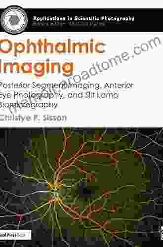
Unlocking the Ocular Secrets: A Comprehensive Guide to Posterior Segment Imaging, Anterior Eye Photography, and Slit Lamp Biomicrography

The human eye is a remarkable organ, capable of capturing and processing visual information with exceptional precision. To fully understand the intricate workings of the eye and diagnose potential disFree Downloads, ophthalmologists rely on a range of imaging techniques that provide detailed insights into its structures and functions. Among these techniques, Posterior Segment Imaging (PSI),Anterior Eye Photography (AEP),and Slit Lamp Biomicrography (SLB) stand out as essential tools in the ophthalmologist's armamentarium.
5 out of 5
| Language | : | English |
| File size | : | 31107 KB |
| Text-to-Speech | : | Enabled |
| Enhanced typesetting | : | Enabled |
| Screen Reader | : | Supported |
| Print length | : | 386 pages |
This comprehensive guide delves into the principles, applications, and clinical significance of these imaging modalities, empowering eye care professionals with the knowledge and skills necessary to effectively diagnose and manage a wide spectrum of ocular conditions.
Posterior Segment Imaging: Unveiling the Inner Workings of the Eye
Posterior Segment Imaging encompasses a suite of techniques used to visualize the posterior segment of the eye, including the retina, optic nerve, choroid, and vitreous humor. These techniques play a crucial role in the diagnosis and management of various retinal disFree Downloads, such as macular degeneration, diabetic retinopathy, and glaucoma.
- Fundus Photography: This non-invasive technique utilizes a specialized camera to capture high-resolution images of the retina and optic nerve. Fundus photography enables the detection of abnormalities in the retinal vasculature, such as hemorrhages and exudates, as well as structural changes indicative of macular degeneration or diabetic retinopathy.
- Optical Coherence Tomography (OCT): OCT is a non-contact imaging technique that generates cross-sectional images of the retina and choroid. This allows ophthalmologists to visualize the retinal layers in unprecedented detail, aiding in the diagnosis of conditions such as macular edema, epiretinal membranes, and choroidal neovascularization.
- Fluorescein Angiography (FA): FA involves the intravenous injection of a fluorescent dye followed by fundus photography. This technique highlights the retinal vasculature, allowing for the detection of vascular abnormalities, leakages, and occlusions. FA is particularly useful in the diagnosis and monitoring of diabetic retinopathy and macular degeneration.
- Indocyanine Green Angiography (ICGA): ICGA is similar to FA but utilizes a different fluorescent dye that binds to proteins in the choroid. This technique provides enhanced visualization of the choroidal vasculature, aiding in the diagnosis of conditions such as polypoidal choroidal vasculopathy and choroidal neovascularization.
Anterior Eye Photography: Capturing the Frontline of Vision
Anterior Eye Photography involves the use of specialized cameras to capture images of the anterior segment of the eye, including the cornea, iris, lens, and anterior chamber. These images are invaluable in the diagnosis and management of a variety of conditions affecting the front of the eye.
- Slit Lamp Photography: Slit lamp photography utilizes a slit lamp biomicroscope to illuminate and magnify the anterior segment of the eye. This technique allows for the capture of high-resolution images of corneal ulcers, scars, and other surface abnormalities.
- Scheimpflug Imaging: Scheimpflug imaging is a non-contact technique that generates three-dimensional images of the cornea. These images provide detailed information about corneal shape, thickness, and curvature, aiding in the diagnosis and management of conditions such as keratoconus and corneal ectasia.
- Anterior Segment Optical Coherence Tomography (AS-OCT): AS-OCT is a non-contact imaging technique that generates cross-sectional images of the anterior segment of the eye. This allows for the visualization of the cornea, iris, and lens in high resolution, facilitating the diagnosis of conditions such as anterior uveitis, iridocyclitis, and lens subluxation.
Slit Lamp Biomicrography: Illuminating the Microscopic World
Slit Lamp Biomicrography is a technique that utilizes a slit lamp biomicroscope to examine the eye under high magnification. This technique allows ophthalmologists to visualize the microscopic structures of the eye, including the cornea, conjunctiva, iris, and lens, in real-time.
Slit Lamp Biomicrography plays a crucial role in the diagnosis and management of a wide range of ocular conditions, including:
- Corneal ulcers and scars
- Conjunctivitis and uveitis
- Cataracts and other lens opacities
- Glaucoma and other anterior chamber abnormalities
Clinical Significance and Applications
Posterior Segment Imaging, Anterior Eye Photography, and Slit Lamp Biomicrography are essential tools in the clinical practice of ophthalmology, providing ophthalmologists with valuable insights into the health and function of the eye. These imaging techniques have revolutionized the diagnosis and management of a wide spectrum of ocular conditions, leading to improved patient outcomes and enhanced quality of life.
- Macular Degeneration: PSI techniques such as fundus photography and OCT play a crucial role in the diagnosis and monitoring of macular degeneration, the leading cause of irreversible vision loss in the elderly. These techniques allow for the detection of early signs of the disease, enabling timely intervention and treatment.
- Diabetic Retinopathy: PSI techniques are essential in the diagnosis and management of diabetic retinopathy, a leading cause of blindness in diabetic patients. Fundus photography, FA, and OCT provide detailed visualization of retinal changes, allowing for the early detection and treatment of this devastating disease.
- Glaucoma: PSI and AEP techniques are invaluable in the diagnosis and monitoring of glaucoma, a condition characterized by increased intraocular pressure and damage to the optic nerve. Fundus photography, OCT, and SLB provide insights into the structural changes associated with glaucoma, aiding in the timely initiation of treatment.
- Corneal DisFree Downloads: AEP techniques such as slit lamp photography and Scheimpflug imaging are essential in the diagnosis and management of corneal disFree Downloads. These techniques provide detailed images of the cornea, allowing for the detection of corneal ulcers, scars, and other abnormalities.
- Cataracts: SLB and AS-OCT are valuable tools in the evaluation of cataracts, a clouding of the lens that can lead to vision impairment. These techniques provide insights into the extent and progression of cataracts, aiding in the decision-making process regarding surgical intervention.
Posterior Segment Imaging, Anterior Eye Photography, and Slit Lamp Biomicrography are indispensable tools in the armamentarium of ophthalmologists, providing invaluable insights into the health and function of the human eye. These imaging techniques have transformed the diagnosis and management of ocular conditions, leading to improved patient outcomes and enhanced quality of life.
By embracing these cutting-edge technologies, eye care professionals can continue to push the boundaries of ophthalmic care, unlocking the secrets of the eye and empowering patients with the gift of clear vision.
5 out of 5
| Language | : | English |
| File size | : | 31107 KB |
| Text-to-Speech | : | Enabled |
| Enhanced typesetting | : | Enabled |
| Screen Reader | : | Supported |
| Print length | : | 386 pages |
Do you want to contribute by writing guest posts on this blog?
Please contact us and send us a resume of previous articles that you have written.
 Book
Book Novel
Novel Page
Page Chapter
 Text
Text Story
Story Genre
Genre Reader
 Library
 Paperback
 E-book
 Magazine
 Newspaper
 Paragraph
 Sentence
 Bookmark
 Shelf
Shelf Glossary
 Bibliography
 Foreword
 Preface
 Synopsis
 Annotation
 Footnote
 Manuscript
 Scroll
 Codex
Codex Tome
Tome Bestseller
 Classics
 Library card
 Narrative
 Biography
 Autobiography
 Memoir
 Reference
 Encyclopedia
 Waka T Brown
 Diana Stout
 Kedar N Prasad
 Steven T Fleming
 Claudio Rivera
 Andrea Mccloud
 Margaret Morrison
 Terry Ryan
 Alex Vin
 Astrid Dumontet
 Evelyn Mcdonnell
 Tim Gorman
 Craig Huffman
 Irwin W Sherman
 Roger Nygard
 Lesley Crossingham
 Carol Crowe Carraco
 Jessica Jones
 Allen C Guelzo
 Gail Thackray
Light bulbAdvertise smarter! Our strategic ad space ensures maximum exposure. Reserve your spot today!
 Bernard PowellEmergency Care and Transportation of the Sick and Injured: A Comprehensive...
 Henry Wadsworth LongfellowBuild Your Own Brick House: A Comprehensive Guide to Constructing a Durable...


 Eugene ScottFollow ·7.4k
 Jacques BellFollow ·6.7k
 Dwayne MitchellFollow ·6.4k
 George HayesFollow ·15.3k
 Camden MitchellFollow ·11.6k
 Paul ReedFollow ·11.4k
 Clark BellFollow ·8.2k
 Tennessee WilliamsFollow ·2.9k
 Henry Green
Corrosion and Its Consequences for Reinforced Concrete...
Corrosion is a major threat to reinforced...
 James Gray
Discover the Enigmatic World of Pascin in "Pascin Mega...
Immerse Yourself in the...
 George R.R. Martin
Unlocking the Power of Nature: Delve into the Bioactive...
In a world increasingly...
 Julian Powell
Master the Art of Apple Watch App Development: A...
Unlock the Potential of Apple Watch Apps In...
 Jaylen Mitchell
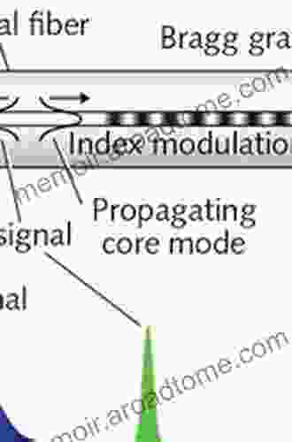
Plastic Optical Fiber Sensors: A Comprehensive Guide to...
In the rapidly evolving landscape of...
 Truman Capote
Unlock the Secrets of Language Creation: Dive into...
The realm of computer science...
5 out of 5
| Language | : | English |
| File size | : | 31107 KB |
| Text-to-Speech | : | Enabled |
| Enhanced typesetting | : | Enabled |
| Screen Reader | : | Supported |
| Print length | : | 386 pages |
